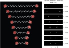
If you want to measure an everyday object, you might use a ruler – a piece of material with a fixed length and regularly-marked divisions. Thanks to a new device called a PicoRuler, the same measurement principle can now be applied to tiny objects such as cells and molecules. Developed by researchers at Julius-Maximilians Universität (JMU) Würzburg in Germany, the miniscule measuring stick works in biological environments and could be used to test the ability of super-resolution microscopy techniques to image objects less than 10 nm long.
Super-resolution microscopy based on fluorescence imaging has developed rapidly over the past 20 years. It is now routine for such methods to resolve structures as small as a few nanometres – far below the diffraction limit for conventional visible-light microscopy.
To push these techniques further, researchers need reference structures to calibrate their microscopes’ performance. The main calibration method currently in use relies on artificial DNA origami structures. These can be synthesized to carry several fluorophores at well-defined positions less than 10 nm apart, allowing them to act like rulers for sub-10 nm imaging. The problem is that DNA origami is highly negatively charged and thus cannot be used in real-world biological cellular imaging media.
Clicking into place
Led by biotechnologists Markus Sauer and Gerti Beliu, the JMU team developed a biocompatible alternative based on a three-part protein called proliferating cell nuclear antigen (PCNA). By introducing synthetic amino acids onto this protein at precisely defined positions spaced 6 nm apart, they made it possible for fluorescent dye molecules to chemically “click” onto it in an efficient way. This new structure allowed them to test the resolution of a technique known as DNA-based points accumulation for imaging in nanoscale topography (DNA-PAINT) down to 6 nm. Sauer says it could also be important for other techniques such as direct stochastic optical reconstruction microscopy (dSTORM), MINFLUX or MINSTED.
“These advanced microscopy techniques can achieve spatial resolutions in the range of a few nanometres, and the new ruler will serve as a calibration tool to verify and enhance their accuracy,” he says.
Exploring cell structure from within
The researchers are now looking to optimize their ruler for use in various biological environments, including living cells. Another direction for development, Sauer says, could be to deliver PicoRulers directly into the cells themselves through techniques like microinjection or functionalization with cell-penetrating peptides. The devices could thus be used to explore the structure of a cell from within, gaining knowledge that might advance cellular biology and bring a better understanding of diseases and pathways to drug development.

Super-resolution microscopy reveals coronavirus-replicating machinery
“Our team is also focusing on expanding the range of biomolecules that can be used as PicoRulers,” Sauer tells Physics World. “To this end we will be looking into different proteins and other biological complexes. We are convinced that the development of our PicoRuler marks a significant step forward in the field of super-resolution microscopy, offering a valuable tool for exploring cellular and molecular structures at unprecedented resolutions.”
The PicoRuler is described in Advanced Materials.


