X-ray detectors play a key role in a wide range of medical applications, including diagnostic imaging, radiotherapy dosimetry and personal radiation protection. Many of these applications require large-area detectors that can flexibly conform to curved surfaces. But most commercial X-ray detectors are stiff, power-hungry and expensive to fabricate into large areas.
One alternative is organic semiconductors, which can be used to create large-area optoelectronic devices via environmentally friendly, low-cost manufacturing techniques. Organic materials, however, exhibit low X-ray attenuation, resulting in detectors with low sensitivity. A team headed up at the University of Surrey’s Advanced Technology Institute aims to solve this problem. By adding small amounts of high-Z elements to an organic semiconductor, the researchers created organic X-ray detectors with high sensitivity and high flexibility.
“This new material is flexible, low-cost and sensitive. But what’s exciting is that this material is tissue equivalent,” explains first author Prabodhi Nanayakkara in a press statement. “This paves the way for live dosimetry, which just isn’t possible with current technology.”
Heavy heteroatoms
To fabricate the new X-ray absorber material, the researchers modified the polymer chain of an organic semiconductor with high-Z selenium heteroatoms to create a p-type polymer, P3HSe, and blended this with an n-type fullerene derivative, PC70BM. They created the X-ray detector on a glass substrate using a 55 µm-thick absorber layer.
Nanayakkara and colleagues evaluated the response characteristics of the new detector, comparing its performance to that of their previous curved X-ray detector candidate, made using bismuth oxide nanoparticles integrated into an organic bulk heterojunction (NP-BHJ).
They first measured the dark current, which determines a detector’s limit of detection, signal-to-noise ratio and dynamic range – crucial parameters in dosimetry and medical imaging. The P3HSe:PC70BM detectors demonstrated an ultralow dark current of 0.32 pA/mm2 under an applied bias of −10 V, well within the industrial standard of 10 pA/mm2 and comparable to that of the NP-BHJ detectors. The researchers point out that these two X-ray detectors display the lowest dark currents reported to date of all organic, hybrid and perovskite detectors in the literature.
To evaluate the sensitivity of the detectors, the team exposed them to various X-ray sources. When exposed to 70, 100, 150 and 220 kVp X-ray radiation, the P3HSe:PC70BM detectors exhibited sensitivities of 22.6, 540, 600 and 550 nC/Gy/cm2, respectively. Again, these values are similar to those observed from the NP-BHJ detectors.
The heteroatom-based detectors also displayed excellent dose and dose rate linearity, as well as high reproducibility under repeated X-ray exposure. The researchers note that “despite the relatively low thickness of these absorbers, P3HSe:PC70BM and NP-BHJ detectors display a satisfactory performance compared to more established, state-of-the-art detector technologies”.
The new detectors also exhibited long-term stability. After 12 months storage in nitrogen in the dark, they showed a slight increase in dark current (although remaining well within industrial standards) and no noticeable variation in X-ray photocurrent response. Repeated X-ray exposures to a cumulative dose of 100 Gy did not degrade detector performance.
Creating the curves
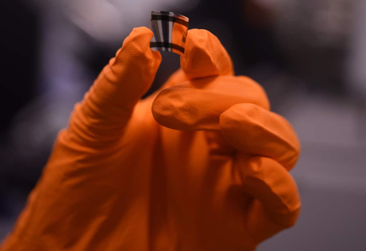
Next, the researchers used the new material to fabricate curved X-ray detectors. As the P3HSe:PC70BM films exhibited similar stiffness and hardness to NP-BHJ films, they employed the same 75 µm-thick polyimide films previously used with the NP-BHJ system as flexible substrates.
To assess the response while deformed, the team exposed P3HSe:PC70BM detectors with bending radii from 11.5 to 2 mm to 40 kVp X-rays. At a bending radius of 11.5 mm, the detectors had a sensitivity of 0.1 µC/Gy/cm2 and a dark current as low as 0.03 pA/mm2 when biased at −10 V. Up to a threshold radius of 3.5 mm, the detectors did not display significant change in sensitivity, but beyond this limit, the photocurrent reduced considerably from the sensitivity in pristine condition.
Examining the performance before, during and after bending the detector to a radius of 2 mm revealed that its sensitivity decreased by about 20% during bending, then recovered to near its initial value after relaxation.
Finally, the researchers assessed the mechanical robustness of the device. After 100 bending cycles down to a radius of 2 mm, the curved detectors showed no signs of mechanical failure and less than 1.2% variation in sensitivity. The team concludes that heteroatom incorporation provides a successful strategy for creating high-performance X-ray detectors based on organic semiconductors.

Could curved X-ray detectors herald the next evolution in medical imaging?
“This is another route to making flexible X-ray detectors, staying firmly only with organic materials,” Ravi Silva, director of the Advanced Technology Institute, tells Physics World. “Both systems show X-ray detectors with broadband high sensitivity and ultralow dark current response. This system based only on organic semiconductors fully preserves the tissue equivalence and will give highly accurate mapping of the X-ray signal, which may not need post-processing so can be used with AI for early detection of tumours.”
Silva adds that this new technology could be used in a variety of settings, including radiotherapy, scanning historical artefacts and in security scanners. “The University of Surrey, together with its spin out SilverRay, continues to lead the way in flexible X-ray detectors – we’re pleased to see the technology shows real promise for a range of uses,” he says. “Mammography and real-time therapeutics including surgery will also be possible. SilverRay is looking at some of these possibilities as we speak.”
The flexible organic X-ray detector is described in Advanced Science.


