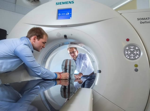
Together with its clinical partners in Europe and the US, Siemens Healthineers is focusing on translational research and technology innovation to drive continuous improvement in the planning, delivery and management of proton therapy for cancer patients

From collaboration comes innovation: that’s certainly the mantra of an ambitious, multicentre German R&D initiative which is leveraging the cross-disciplinary expertise of academic researchers, clinicians and industry to deliver game-changing advances in the radiation oncology clinic – enhancing the accuracy, safety and tissue-sparing capability of proton therapy systems in the process. The breakthroughs in question stem from the clinical deployment of dual-energy CT (DECT) for proton treatment planning and the application of so-called DirectSPR software1,2 for the more accurate prediction of proton range in the patient’s body. All part of a unifying vision – think translational research meets clinical application – to reimagine the planning, delivery and management of proton treatments tailored to the unique requirements of individual cancer patients.
The clinical roll-out of DirectSPR is being pioneered by a translational team headed by Christian Richter at the Dresden-based OncoRay. The OncoRay researchers work closely on algorithm development and validation with their colleagues at the German Cancer Research Center (DKFZ) in Heidelberg – the two institutes together form the National Center for Radiation Research in Oncology (NCRO) – while industrial support comes from the Cancer Therapy Business Line at Siemens Healthineers.
OncoRay itself is a publicly funded research centre that focuses on translating new technologies and treatment methods in radiation oncology into clinical application, pooling the strengths of its three founding institutions: Carl Gustav Carus University Hospital Dresden, Technische Universität Dresden and the Helmholtz-Zentrum Dresden-Rossendorf. “While OncoRay’s principal driver is to improve treatment efficacy and patient outcomes,” explains Richter, “we also seek partnerships with leading medical technology manufacturers to ensure that our R&D reaches the wider radiation oncology community – with DECT-based DirectSPR a notable success story in both respects.”
Innovation unpacked
If that’s the back-story, what of the specifics underpinning this new paradigm in proton treatment planning – and in particular, the basis for the more accurate predictions of proton range and stopping behaviour in different tissue types? Key to success is the innovative application of DECT technology and its spectral imaging capability to the prediction of proton range for treatment planning. Unlike conventional CT, this imaging modality involves the acquisition of two separate X-ray energy spectra – an approach that, in turn, allows the characterization of tissues exhibiting different attenuation properties at different energies (though, crucially, without exposing patients to a higher X-ray dose).
DirectSPR, meanwhile, is an algorithm for post-processing of DECT images – and specifically the determination of the individual stopping behaviour of protons in different tissue types. At the same time, DirectSPR maximizes the quantitative quality of the stopping information by using sophisticated noise reduction and by tailoring the application to the scanned body region of interest. In this way, DirectSPR makes it possible for medical physicists to better resolve differences in tissue composition across a given patient cohort – as well as in the same patient – and to take these differences into account during treatment planning. (For completeness, SPR is the stopping-power ratio and expresses the energy loss of the protons versus distance travelled through the patient relative to the energy loss in water.)

In Spring 2019, Richter and his colleagues at OncoRay became the first team to deploy DECT-based DirectSPR as part of the routine clinical workflow for proton therapy – a key step in the improvement of treatment planning of static tumour sites at University Proton Therapy Dresden (part of Carl Gustav Carus University Hospital). Since then, for more than 300 patients, DECT-based DirectSPR has enabled the Dresden clinic – on a sustained and repeatable basis – to reduce the volume of irradiated healthy tissue surrounding the target volume (the safety margin) by approximately 35% for prostate cancer and brain tumour treatments.
Clinical impact, of course, is defined along multiple coordinates, not least the accuracy of SPR calculations and related proton-range predictions – both of which dictate the safety margin around the tumour and, by extension, the degree of tissue sparing. Thanks to the clinical introduction of DECT-based DirectSPR, OncoRay has realized significant reductions in the uncertainty of its proton-range calculations (now <2%) versus traditional CT-based planning approaches (where uncertainties have been set at 3.5% of total proton range for the past 30 years). The result: proton-range reductions on average of 3.6 mm for pelvic treatments and 2.6 mm for treatments in the head. “For individual brain tumour treatments,” says Richter, “we are therefore able to reduce dose to critical structures like the brain stem by 16% as well as the optic chiasm and optic nerve by 7%, with 4% reduction overall in mean dose to the brain as shown in a representative patient case. That’s relevant progress in minimizing the patient’s risk of post-treatment side-effects.”
Translation in action
The timeline for translation of DirectSPR into a clinical product can be traced back to early 2015, when scientists from OncoRay and the DKFZ began work on the development and optimization of the DirectSPR approach. Their aim: to minimize the uncertainty associated with proton-range predictions in the patient’s tissues and thereby enhance targeting accuracy and dose distribution accuracy for proton treatments. This four-year, NCRO-funded project – carried out in close collaboration with Siemens Healthineers – began with the introduction of DECT for clinical proton therapy planning at University Proton Therapy Dresden in 2015 – a world-first that made it possible to retrospectively evaluate the DirectSPR approach on a growing database of therapeutic DECT patient scans (rather than just using phantoms or computer simulation).
“Even in the formative stages of DirectSPR algorithm development, OncoRay and DKFZ were focused on translational outcomes,” says Richter. The definition of a sophisticated algorithm early on – and especially the subsequent focus on comprehensive stepwise validation in different scenarios (including an anthropomorphic phantom and biological tissues) – was fundamental to successful translation. “This convinced us, the community and the responsible clinicians of the superiority of the approach,” adds Richter. “Siemens Healthineers came on board in 2016, with the early conversations building trust and understanding around our mutual clinical and research goals.”
It was at this point that the collaboration shifted gears into the preclinical product development phase, with OncoRay, DKFZ and Siemens Healthineers working together on optimization and calibration of the DirectSPR algorithm. With the commercial launch of DirectSPR software in spring 2019, Siemens Healthineers now ensures compatibility of DirectSPR across its full portfolio of DECT scanners with dedicated calibrations for each CT model.
The clinical roadmap
In terms of next steps, Richter and colleagues at OncoRay – as well as their industry counterparts at Siemens Healthineers – are focused on accelerating clinical acceptance of DirectSPR within the proton therapy community (see “Proton perspectives: the DirectSPR opportunity”). OncoRay, for its part, maintains bilateral collaborations with many European proton therapy centres and is also active within the European Particle Therapy Network, a subgroup of the European Society for Radiotherapy and Oncology (ESTRO). “A consensus paper is in preparation to define best-practice guidelines and standardization for the clinical implementation of DirectSPR in the proton therapy clinic,” explains Richter. That document, he hopes, will be brought forward during discussions at the upcoming ESTRO workshop on Clinical Translation of CT Innovations in Radiation Oncology.
Meanwhile, the OncoRay and DKFZ teams are already working with Siemens Healthineers to explore new translational pathways, including the use of DECT-based DirectSPR for proton treatment planning on moving targets – for example, tumour sites in the lung and abdomen. “Our task at OncoRay and DKFZ is to support further improvement of DirectSPR for next-generation CT scanners in a radiotherapy setting,” concludes Richter.
Proton perspectives: the DECT-based DirectSPR opportunity
There’s growing clinical appetite to deploy DirectSPR as a core component of the proton therapy workflow. Here Physics World talks to medical physicists at some of the early-adopting proton facilities to assess progress, next steps and anticipated clinical upsides.
Tianyu Zhao, Washington University School of Medicine in St Louis, Missouri
We are recruiting patients into a National Institutes of Health (NIH)-sponsored study, collecting data using DECT scanners to improve estimates of tissue composition accuracy for tumour sites and surrounding organs. The goal: to reduce the discrepancy between the actual dose delivered versus the planned dose in proton therapy. We plan to quantify the accuracy of DirectSPR for mass density and proton SPR predictions by using phantoms scanned with clinical protocols. This site-specific accuracy information will be used to adjust the uncertainty setting in robustness optimization for proton patients scanned with DECT. Ultimately, the hope is that our work will reduce the range uncertainty for protons and lead to better planning quality and fewer dose prediction errors in proton therapy.
Ming Yang, MD Anderson Cancer Center, Houston, Texas
We are currently upgrading our institutional syngo.via server (from Siemens Healthineers) to VB40 – a prerequisite for implementing DirectSPR. Upon completion, we will start the commissioning process as soon as DirectSPR is available to us. Initial priorities will be to test DirectSPR’s compatibility with our existing treatment planning system and to evaluate the uncertainties associated with the software’s estimation of proton stopping power. Personally, I believe DirectSPR will benefit our patient population in multiple ways. One immediate impact will be a reduction in normal tissues receiving high dose close to prescription dose – so a reduction in normal-tissue toxicity. A smaller range uncertainty parameter will also make it easier to achieve a robust treatment plan meeting the planning objectives. Finally, the reduced range uncertainty might also give us more choices of beam angle to take full advantage of the Bragg peak.
Benjamin Ackermann, Heidelberg Ion Beam Therapy Center (HIT), and Friderike Longarino, Heidelberg University Hospital
HIT completed clinical validation of DirectSPR using a free-of-charge test licence – a convenient and low-risk option for clinics interested in finding out more. The validation process included a complete radiotherapy workflow and dosimetric tests in anthropomorphic phantoms, consisting of a multitude of different tissue-equivalent materials. We are now installing the purchased software licence and looking forward to performing the first patient scans using DirectSPR, starting with body regions like the head and pelvis which exhibit little or no intrafractional movement. With DECT scans and DirectSPR image processing, we expect to achieve better SPR predictions and, in turn, better range prediction in ion beam therapy compared to SECT. Another anticipated benefit is a reduction in safety margins for enhanced sparing of healthy tissue from unwanted irradiation.
Sina Mossahebi, Maryland Proton Treatment Center, US
In our initial DirectSPR study, we aim to quantify dose-calculation discrepancies between conventional SECT and DECT in vivo and on patient plans, with the objective of reducing range uncertainty in the planning of intensity-modulated proton therapy (IMPT). Both SECT and DECT scans are acquired from the same patient and used to evaluate dosimetric differences between treatment plans generated on each image set. The primary goal of the study is to quantify and compare the target coverage and dose to organs at risk (OARs) as well as the plan robustness between SECT and DECT scans obtained for patients undergoing IMPT. In addition, we will validate SECT and DECT dose calculations using patient-specific QA, while exploring practical advantages of DECT with respect to target delineation and implants.
Gabriel Fonseca and Frank Verhaegen, Maastro Clinic, The Netherlands
Our evaluation of DirectSPR is proceeding along multiple fronts. Through extensive phantom testing, we have investigated the SPR information used in the dose calculations by the TPS, comparing SPR predictions obtained via the DECT-based DirectSPR and SECT approaches. The former is more accurate, exhibiting differences of less than half of those obtained by SECT. Another focus is the calibration procedure, which will likely require a range of phantom sizes to match variations in patient size – i.e. extra work in the clinic. Although DirectSPR can provide more accurate SPR values, the treatment plan uncertainties depend on several factors. As such, the Maastro team is evaluating the effect of improved SPR maps on robust optimized plans for a cohort of more than 50 patients. In summary: while we expect DirectSPR to yield improved proton range accuracy from 1 mm up to several mm (for some deep-seated tumours), it’s clear that all sources of uncertainty and risk need to be quantified thoroughly prior to routine clinical deployment. Long term, strict QA procedures will need to underpin the clinical workflow.
Ole Nørrevang, Danish Centre for Particle Therapy, Aarhus University Hospital, Denmark
We’re taking a phased approach to the roll-out of DECT-based DirectSPR here in Aarhus. We started out by implementing virtual monoenergetic images and optimizing the use for both organ and target delineation and stopping-power estimation. With DirectSPR software now available to us, our investigations are proceeding on three fronts: validation of the SPR values produced by DirectSPR; studying the effect of reduced range uncertainty in terms of reduced dose to OARs in the brain; and understanding the workflow, patient safety and transition issues associated with the use of SPR images in the treatment planning of brain tumours. Our focus for now is on the treatment of brain tumours, because we can exclude the effects of motion artefacts in the DECT image. Over time, though, we will develop plans to apply DirectSPR for other treatment sites.
Koen Salvo, PARTICLE Proton Therapy Center Leuven, Belgium
We are currently in the process of defining our DECT simulation protocols, with the adult protocols complete and conversations in progress with Siemens Healthineers on the child protocols. Once those protocols are finalized, we will activate our DirectSPR software and validate it using proton radiography on animal phantoms. With DirectSPR, we hope to calculate the proton range more accurately. If so, we can reduce our target margins and, in turn, reduce the dose to OARs near the target – a significant advance when treating chordomas near the brain stem.
End note: the statements by Siemens Healthineers’ customers described herein are based on results that were achieved in the customer’s unique setting. Because there is no “typical” hospital or laboratory and many variables exist (e.g. hospital size, sample mix, case mix, level of IT and/or automation adoption), there can be no guarantee that other customers will achieve the same results. 1) DirectSPR is an optional feature. 2) DirectSPR calculations require Dual Spiral Dual Energy, Twin Spiral Dual Energy or Dual Source acquisition; TwinBeam Dual Energy data are not supported for DirectSPR calculations. More information on DirectSPR and its requirements can be found here.


